Home > Laser surgery / Removal of tumors / Removal of hemangiomas > Treatment of hemangiomas of the tongue
Tongue hemangioma develops in people of different ages. The appearance and symptoms of such a tumor depend on the type of tumor.
Hemangioma of the tongue is a benign formation that grows very slowly. One thing about hemangioma is that it can grow very quickly after a period of slow development. The tongue disease affects young children, adults and elderly patients. The method of treating the disease is removal.
Consultation on the day of the procedure is free
Symptoms and causes of formation
Hemangioma of the tongue can be:
- cavernous;
- capillary.
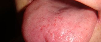
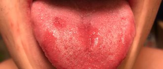
A simple capillary hemangioma grows in breadth without affecting the tissue. It consists of capillary tissue and is often affected during conversation or eating. Externally, the tumor looks like a spot or bruise. The formation must be removed - damage to the hemangioma can lead to infection.
Cavernous hemangioma of the tongue penetrates deep into the tissues; it protrudes noticeably above the surface of the tongue. This formation consists of many vessels and causes serious discomfort. The tongue may lose its mobility and increase in size due to the tumor.
The causes of hemangiomas have not been fully established. In children they occur mainly due to the growth of vascular tissue.
Causes and mechanisms
The development of lumps on the roof of the mouth always indicates pathology. However, they can have different origins. Among the diseases that have this manifestation may be:
- Benign tumors (myxoma, angioma, fibroma, papilloma, osteoma, cyst).
- Viral diseases (herpes).
- Exostoses (bone protrusions).
- Lichen planus.
- Syphilis (chancre or gumma).
- Leukoplakia of the oral cavity.
- Oncological processes (cancer).
Thus, tubercles on the mucous membrane are most often of inflammatory or proliferative origin. Various factors contribute to the appearance of such changes:
- Mechanical trauma (with dentures, after operations).
- Burns (hot food, chemicals).
- Bad habits (smoking, alcohol, drugs).
- Congenital anomalies, etc.
Most often we are talking about local disorders in the oral cavity, but disorders of a systemic nature cannot be ruled out, against the background of which growths form on the palate. And each case requires an individual approach to diagnosis.
Pathological formations on the oral mucosa can arise due to various conditions that require timely detection.
Danger of hemangiomas
If a hemangioma appears on the tongue, you should urgently consult a doctor. A benign tumor can be dangerous. Capillary and cavernous types of hemangiomas are damaged during the absorption of food. With cavernous hemangiomas, bleeding is possible. This threatens the penetration of infections.
A simple capillary hemangioma may not cause discomfort. Some patients don't even feel it. But this does not mean that the disease can be ignored. The unexpected can happen at any moment. There are a lot of bacteria on the mucous membrane of the tongue, and it is only a matter of time before infection penetrates into the damaged tumor. Do not delay contacting a specialist.
At the clinic, a patient with a hemangioma will be prescribed treatment. The tumor must be removed. When removing, several methods are used:
- laser;
- surgical method;
- cryotherapy;
- removal by radio wave equipment.
Laser is the most popular method for removing hemangiomas. However, this type of treatment is not always possible. In some cases, doctors are able to solve the problem with a simpler surgical method.
Symptoms
You can find out about the reasons for what is happening based on an analysis of clinical manifestations. Any diagnostic program begins with a medical examination, regardless of the nature of the pathology. First, a survey and examination are carried out, followed by physical methods (for example, palpation), and the information obtained, if necessary, is supplemented with laboratory and instrumental studies.
Benign tumors
Growths on the palate are often benign. The most common tumors of the oral cavity are hemangiomas. They can be capillary, cavernous and mixed. The tubercle on the mucous membrane has a soft consistency and a red-bluish color, and collapses when pressed. Very often, hemangiomas are injured, which leads to bleeding.
Fibromas consisting of connective tissue often form on the palate. Their shape is round or oval, the consistency is densely elastic. The tumor is not fused with the surrounding tissues and is surrounded by a capsule, often growing on a thin stalk. The color of fibroma does not differ from the normal mucous membrane. Often, a fibroma can transform into a myxoma, a mucous soft tumor of a whitish color.
Cost of hemangiomas removal
| Laser tumor removal | Prices, rub. |
| Laser removal of papillomas, single warts - Cat. I. difficulties | 1200 |
| Laser removal of papillomas, multiple warts - Cat. I. difficulties | 350 |
| Laser removal of moles, papillomas, warts - Cat. II. difficulties | 700 |
| Laser removal of moles, papillomas, warts - Cat. III. difficulties | 1500 |
| Laser removal of moles, papillomas, warts - IV category. difficulties | 3000 |
| Laser removal of moles, papillomas, warts - Cat. V. difficulties | 4500 |
| Laser removal of moles, papillomas, warts - Cat. VI. difficulties | 6100 |
| CO2 Laser callus removal (per unit) | 6100 |
| Removal of atheroma, lipoma, fibroma, xanthelasma with laser - Cat. I. difficulties | 6 900 |
| Removal of atheroma, basal cell carcinoma, lipoma, fibroma, xanthelasma with laser - Category II. difficulties | 9 400 |
| Removal of atheroma, basal cell carcinoma, lipoma, fibroma, xanthelasma with laser - Cat. III. difficulties | 16 900 |
| Removal of atheroma, basal cell carcinoma, lipoma, fibroma, xanthelasma with laser - IV category. difficulties | 22 400 |
| Removal of atheroma, basal cell carcinoma, lipoma, fibroma, xanthelasma with laser - Cat. V. difficulties | 33 400 |
Sign up for laser removal of tongue hemangioma
- Full name
- Telephone
Why are red moles dangerous?
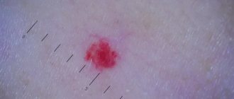
If a red mole does not bother its owner and does not change its external characteristics, it can be argued that such a formation does not pose a significant threat to the health and life of the patient. At the same time, you should get professional medical advice and resort to modern therapeutic methods if you have the following manifestations:
- The red mole grows or changes its shape;
- The patient experiences pain, itching or burning in the area where the pigmented formation is located;
- The mole is bleeding;
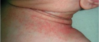
- Superficial structures or ulcerations appear;
- There are more than 6 small red dots in one area of the body.
All these symptoms may indicate the development of an oncological process. In addition, the category of dangerous moles includes angiomas located in places where they can be easily injured when wearing clothes, shoes, jewelry, etc. Mechanical damage to such a formation can provoke the appearance of new red moles on the body, activate malignant transformation and leave scars or scars on the skin.
Removal methods
When treating a tumor, the patient is prescribed removal. The method of removal depends on the complexity of the disease. If there is no risk of damaging surrounding tissues, then a surgical method is used. This treatment is used if the tumor has not penetrated inside. In this case, the surgeon will be able to remove all the affected cells.
Surgical treatment is not suitable if the tumor has penetrated deep into the tissue. There is a risk of damaging the tongue and not removing damaged cells completely. In difficult cases, the radio wave method is prescribed. It is used to treat cavernous hemangiomas. Affected cells are removed at ultra-high temperatures.
Treatment with ultra-low temperatures - cryotherapy - is also possible. During treatment, applications are applied to the tongue, which freeze the affected cells. The tumor tissues die and separate.
Causes of red moles on the body
By its nature, a red mole is a cluster of vessels and capillaries, which normally perform the function of oxygen and nutritional supply to the epidermal structures. With the development of any internal pathological process or under the influence of exogenous factors, small vessels can connect, forming a kind of bundle, which in medicine is often called an angioma.
When asked by a patient regarding small red moles, “What is this?”, the specialist will most often answer – angioma. Typically, these pigmented formations are formed during the period of active growth of the body, that is, in early childhood, which is associated with a serious transformation of the human vascular system at this age.
A distinctive feature of red pigmented nevi is the loss of color intensity of the formation when pressing on it, which is associated with a vascular reaction. In addition, red moles normally do not hurt or itch, and do not change their shape and size over time.
The following etiotropic factors may be the causes of angiomas in adult patients:
- Excessive sun exposure (excessive ultraviolet radiation);
- Hormonal imbalance (puberty, pregnancy, menopause, etc.);
- Pathologies of the gastrointestinal tract, especially the pancreas;
- Injuries and mechanical damage to the epidermal integument;
- Diseases of the cardiovascular system;
- Violation of the formation or destruction of pigmented cells.
Today, doctors do not give a clear answer regarding the causes of red moles on the body, however, the factors listed above can become a provoking condition for their appearance.
Laser treatment
The easiest way to remove a hemangioma is with a laser. This treatment method has several advantages:
- no risk of infection;
- This is a non-contact method of therapy;
- tumor cells are completely removed;
- the procedure is painless;
- It is possible to cure even cavernous tumors;
- the wound heals quickly.
Laser treatment does not take much time. The procedure uses a laser with a short beam length. Removal does not cause serious discomfort to the patient. The laser is absolutely safe, the procedure does not lead to side effects. After treatment of hemangioma of the tongue, no bleeding is observed.
Complete laser removal leads to rapid restoration of healthy tissue. The tongue heals and the patient returns to normal life. A special laser is used in clinics where they offer the procedure for removing hemangioma in this way.
Removal of tumors at Lazmed Clinic
Types of red moles
The appearance of red moles on the body can be accompanied by various visual characteristics of the defect. Such epidermal formations can be either flat, forming in the deep dermal layers, or have a superficial nature, protruding above the skin. Moreover, according to the nature of the visual picture, all angiomas are usually divided into two groups:
- Spot. The pigmented area has clear red borders and is a point at which a collection of vessels and capillaries is concentrated. Often, punctate angiomas are multiple in nature, manifesting themselves in the form of specific “rashes” on the patient’s skin.
- Star-shaped. Such moles are a collection of tiny thin vessels that are visible through the epidermis and converge at a central point, forming a kind of star. These structures are also associated with diseases such as rosacea.
In addition, a separate group includes especially large red moles on the body - hemangiomas, which usually significantly spoil the patient’s appearance and require cosmetic correction.
Our specialists
- Kiani Ali
Candidate of Medical Sciences, laser medicine specialist, dermatocosmetologist.
Sign up
- Stepanova Inna Igorevna
Candidate of Medical Sciences, maxillofacial surgeon, specialist in laser medicine.
Sign up
- Fedotova Marina Andreevna
Surgeon, dermatocosmetologist, laser medicine specialist
Sign up
- Popovkin Pavel Sergeevich
Surgeon, oncologist, laser medicine specialist.
Sign up
Medical correction of red moles
The doctor, after conducting an initial examination and the necessary diagnostic measures, will make a conclusion regarding the likelihood of malignancy of the red mole. Based on this diagnosis, a strategy for further therapeutic actions is developed. If a red mole does not pose an oncological threat and is located in a closed area of the body, its removal is not necessary.
In cases where the red nevus causes aesthetic or physiological discomfort to the patient, it can be removed using modern hardware techniques, the priority among which is laser destruction. This method of removing red moles guarantees painlessness and safety for the patient, and also has a low level of trauma.
Medical offers its patients the latest expert-class medical equipment that meets the most stringent European standards. Effective therapy, high-quality medical service and affordable prices are the main principles of our clinic.
Find out the cost of the procedure “Removal of tumors”